


EPV app for iPhone and iPad
Developer: Yale
First release : 29 Oct 2015
App size: 40.48 Mb
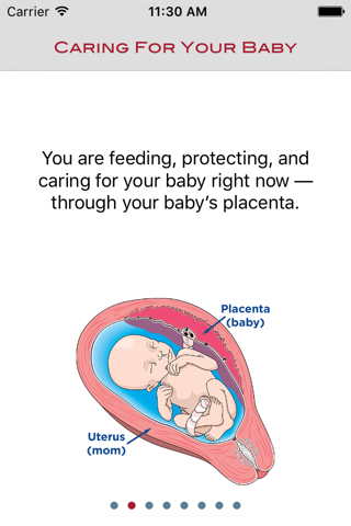
EPV is a ResearchKit app that allows patients to follow the growth of their baby’s placenta (the fetal organ that acts like the roots of a tree, bringing nutrients and oxygen from the mother to the developing fetus). By participating in this study pregnant women can help researchers to understand normal and abnormal placental growth. Once the patient is found to be eligible to join the study and signs the consent form, the Estimated Placental Volume (EPV) can be determined whenever the participant undergoes an ultrasound examination during their pregnancy. At the completion of the pregnancy the patient is asked to report on the pregnancy outcome. No personal information is stored or transmitted to the researchers. All information is transmitted in an encrypted manner and stored as anonymous data on a secure server.
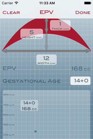
The EPV app calculates the Estimated Placental Volume (EPV) from 2D ultrasounds of the human placenta (1).
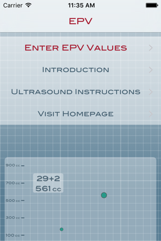
The user interface calculates EPV and then plots the resultant value on a graphical plot. As new values are collected, they are added to the graph. The user can select any of the values to see what the gestational age was at the time of data collection and the EPV for that exam.
Using a simple 2D ultrasound cross-section of the major axis of a placenta from 7 to 45 weeks of gestation the Estimated Placental Volume (EPV) can be calculated from three simple measurements: maximal width, height at maximal width and thickness along the same line as the height. Measurements are recorded in centimeters.
Physicians, nurse midwives, ultrasound technicians and other health care providers can assist the patient use this application to follow placental growth during gestation.
Use of this application should always be done with the assistance of a health care provider. All clinical decisions regarding any patient examined by this method should be made by the patient’s primary care giver for her pregnancy. Any EPV results generated should not be used to make clinical decisions about a patient’s pregnancy or the fetus’ well being.
Key Features:
- Enter the gestational age in weeks and days
- Easily enter width, height and thickness of the placenta—calculates EPV immediately using mathematical modeling
- EPV result is immediately plotted on a graphical plot. Additional EPV data points are plotted on the same graph so the patient and healthcare providers can see the progress of EPV throughout the pregnancy
1. Azpurua HJ, Funai EF, Coraluzzi L, Sasson I, Doherty L, Kliman M, Kliman HJ.
(2010) Determination of placental weight using two-dimensional sonography and volumetric mathematic modeling, Am J Perinatology, 27:151-155.